Go to Rehab Plans
Back to Conditions Pure FCP Service Expert FCPs for your Primary Care Network

Diastasis Recti Abdominis (DRA)

What is diastasis recti abdominis?

  • This is a common condition during and following pregnancy and is caused when the abdominal muscles are overstretched causing a separation in the rectus abdominus (the 6-pack muscles).

How common is diastasis recti abdominis?

  • It is extremely common for a woman to experience a diastasis recti abdominis of some degree during or after pregnancy (10).
  • It can also affect men or women with weakened abdominal muscles and excessive abdominal pressure/rapid weight gain/weight gain (3).

Should I worry?

  • No.
  • Diastasis recti are not linked to any serious pathology (11).
  • More often pregnancy-related diastasis recti (4 fingers and below) will improve within 4 – 6 weeks post-delivery; sometimes it needs additional input from a physiotherapist to aid recovery (12).
  • If your diastasis recti persist and are not resolving, you should seek professional help from a physiotherapist on how best to address this (11). If you have any additional symptoms relating to bladder or bowel function you should contact your GP first (12).

Who is most likely to suffer from diastasis recti abdominis?

  • This condition is mainly associated with pregnancy (11).
  • It can also affect premature babies due to the underdevelopment of abdominal muscles (3).
  • In males, it is linked with an increase in age, weight fluctuations, weightlifting and inherited abdominal weakness (1).

What are the common symptoms?

  • A feeling of a dip in-between the abdominal muscles above or below the belly button or both.
  • Feeling weak through the midsection.
  • Doming or tenting of the middle of the stomach. This can occur during activities such as lifting, rolling over in bed or certain exercises.

What can I do?

  • Exercise during pregnancy might help to reduce your risk of developing diastasis recti (5, 12).
  • Weight management.
  • Pelvic floor strengthening.
  • Post-pregnancy-specific physiotherapy exercises to help strengthen the core (2).

How long will it take to recover?

  • In most cases, diastasis recti heal on its own between the postpartum period (the period following childbirth) of 6 weeks to 3 months (1).
  • It may persist for some time after giving birth; specific exercises can be performed to improve the condition in these cases (12).

1. Introduction

During pregnancy, abdominal muscles stretch to allow the baby room to grow (12). Diastasis recti is a condition in which the abdominal muscles overstretch causing a separation in the rectus abdominus (the 6-pack muscles) which are connected by the linea alba (a thin connective tissue) (12). Commonly separation occurs at the belly button region but it can be anywhere between the xyphoid process (bottom of chest plate) to the pubic bone (10). Usually, there is no pain present in the abdominals. However, as a result, women often report weakness, a mound or ‘flabbiness’ in the affected area (11). Secondary problems can arise such as the low back, hip and pelvic pain (10). Sometimes the condition is referred to as DRAM (divarication of rectus abdominis muscle).

It can also occur in males with links to increasing age, activities and genetic factors. In infants or newborns, it can be inherited or due to the under-development of the muscles in babies born prematurely (1).

pure

2. Signs & Symptoms

Diastasis recti is usually painless in the abdominals, but other symptoms include (3):

3. Causes

In the past, it was thought that body mass index (BMI), weight gain during pregnancy, the weight of the baby and maternal age were considered risk factors. However, a recent study in 2015, found no significant correlation between these factors and developing the condition (1).

During pregnancy, diastasis recti usually resolves spontaneously but can persist postpartum, in some cases for 6 months. In some case where the separation has been significant (6 fingers and above), it may take slightly longer. Assistance with physiotherapy exercises can aid the recovery of the abdominal muscles (10).

pure

4. Risk Factors

This is not an exhaustive list. These factors could increase the likelihood of someone developing diastasis recti. It does not mean everyone with these risk factors will develop symptoms.

5. Prevalence

Diastasis recti is a very common condition following pregnancy. It has been reported to affect between 27%-100% in late pregnancy and 30%-60% of women postpartum (8). Another study has estimated that it occurs in 52% of first pregnancies at 4-6 weeks postpartum and 39% at 6 months (1). Umbilical hernias are less common and it has been found that the incidence is less than 0.1% in pregnant women (9).

6. Assessment & Diagnosis

Musculoskeletal physiotherapists and other appropriately qualified healthcare professionals can provide you with a diagnosis by obtaining a detailed history of your symptoms. A series of physical tests might be performed as part of your assessment to rule out other potentially involved structures and gain a greater understanding of your physical abilities to help facilitate accurate diagnosis.

Your treating clinician will want to know how your condition affects you day-to-day so that treatment can be tailored to your needs and personalised goals can be established. Intermittent reassessment will ascertain if you are making progress towards your goals and will allow appropriate adjustments to your treatment.

Real-time ultrasound imaging can be an accurate way of measuring the gap between abdominal muscles. However, often a healthcare professional can conduct reliable tests in the clinic without the need for a scan (7). Diastasis recti is likely to be confirmed if there is evidence of the following:

  • A palpable gap of more than two and a half finger-widths when performing a curl up.
  • Gapping that does not shrink as abdominals are tightened by the patient.
  • Presence of a small protruding mound at the midline.
  • On lying, a dip in the abdominals/or doming on exertion/stress of the abdominals.

7. Self-Management

As part of your treatment, your musculoskeletal physiotherapist will help you understand the condition and what needs to be implemented to effectively manage your diastasis recti. This will include activity modification strategies, as well as other useful treatments aimed at reducing complications. Regular adherence to a condition-specific rehabilitation programme is important in the management of this condition. It should be noted that rehabilitation exercises are not always a quick fix, but if adhered to on a consistent basis, weeks to months, over time they have been shown to yield positive outcomes (10).

How to check your ‘DRAM’

Lying on your back with knees bent, relax and place a hand above your belly button, fingers straight pointing towards your stomach. Slow curl your head and shoulders up. See how many tips you can fit between your rectus abdominal muscle; you should be able to feel the borders. If you are having difficulty, then draw in the lower tummy muscles first on lying before you curl up. If you are still having trouble, then you can contact a women’s health physiotherapist to help you.

pure

8. Rehabilitation

Specific physiotherapy exercises directed by a healthcare professional that has a good knowledge of the condition is the first line of intervention. It is important to understand what type of exercises you can do to limit over-straining the abdominal area (2).

Below are three rehabilitation programmes created by our specialist physiotherapists targeted at addressing diastasis recti. In some instances, a one-to-one assessment is appropriate to individually tailor targeted rehabilitation. However, these programmes provide an excellent starting point, as well as clearly highlighting exercise progression.

9. Diastasis Recti Abdominis Rehabilitation Plan

Early Plan

This programme focuses on engaging the stomach muscles to begin the process of regaining strength in the area without placing too much stress on the main stomach muscles. This should not exceed any more than 3/10 on your perceived pain scale.

Early Plan  - Rating

Intermediate Plan

This programme provides a progression to the early programme with the focus remaining on the main stomach muscles but with the exercises becoming more challenging. This should not exceed any more than 3/10 on your perceived pain scale.

Intermediate Plan - Rating

Advanced Plan

The exercises in this programme again increase in difficulty. They aim to assist in the process of returning to normal activity levels after the condition. This should not exceed any more than 3/10 on your perceived pain scale.

Advanced Plan  - Rating

10. Return to Sport/Normal Life

For patients wanting to achieve a high level of function or return to sport, we would encourage a consultation with a physiotherapist as you will likely require further progression beyond the advanced rehabilitation stage.

As part of a comprehensive treatment approach, your musculoskeletal physiotherapist may also use a variety of other pain-relieving treatments for associated complications to support symptom relief and recovery. Whilst recovering, you might benefit from a further assessment to ensure you are making progress and to establish the appropriate progression of treatment.

11. Other Treatment Options

If conservative management of diastasis recti is not beneficial and there is a hernia present, surgery can be considered (9). There are two types of hernia surgery:

  • Open hernia repair surgery – the hernia is repaired through a cut (incision) in the belly. Open surgery is safe and effective and has been done for many years.
  • Laparoscopic hernia repair – a surgeon inserts a thin, lighted scope through a small incision in the belly.

References

  1. da Mota, P.G.F., Pascoal, A.G.B.A., Carita, A.I.A.D. & Bø, K. (2015). Prevalence and risk factors of diastasis recti abdominis from late pregnancy to 6 months postpartum, and relationship with lumbo-pelvic pain. Manual therapy, 20(1), 200-205.
  2. Boissonnault, J.S. and Blaschak, M.J. (1988). Incidence of diastasis recti abdominis during the childbearing year. Physical therapy, 68(7), 1082-1086.
  3. Benjamin, D.R., Van de Water, A.T.M. and Peiris, C.L. (2014). Effects of exercise on diastasis of the rectus abdominis muscle in the antenatal and postnatal periods: a systematic review. Physiotherapy, 100(1), 1-8.
  4. Jessen, M. L., Öberg, S., & Rosenberg, J. (2021). Surgical techniques for repair of abdominal rectus diastasis: a scoping review. Journal of Plastic Surgery and Hand Surgery, 1-7.
  5. Chiarello, C.M., Falzone, L.A., McCaslin, K.E., Patel, M.N. & Ulery, K.R. (2005). The effects of an exercise program on diastasis recti abdominis in pregnant women. Journal of Women’s Health Physical Therapy, 29(1), 11-16.
  6. Coldron, Y., Stokes, M.J., Newham, D.J. and Cook, K. (2008). Postpartum characteristics of rectus abdominis on ultrasound imaging. Manual therapy, 13(2), 112-121.
  7. Qu, E., Wu, J., Zhang, M., Wu, L., Zhang, T., Xu, J. & Zhang, X. (2021). The ultrasound diagnostic criteria for diastasis recti and its correlation with pelvic floor dysfunction in early postpartum women. Quantitative Imaging in Medicine and Surgery, 11(2), 706.
  8. Rett, M.T., Braga, M.D., Bernardes, N.O. & Andrade, S.C. (2009). Prevalence of diastasis of the rectus abdominis muscles immediately postpartum: comparison between primiparae and multiparae. Brazilian Journal of Physical Therapy, 13(4), 275-280.
  9. Kulacoglu, H. (2018). Umbilical hernia repair and pregnancy: before, during, after…. Frontiers in surgery, 5, 1.
  10. Lee, D. & Hodges, P.W. (2016). Behaviour of the linea alba during a curl-up task in diastasis rectus abdominis: an observational study. journal of orthopaedic & sports physical therapy, 46(7), pp.580-589.
  11. Lee, D.G. (2011). The Pelvic Girdle E-Book: An integration of clinical expertise and research. Elsevier Health Sciences.
  12. POGP (2020) ‘Tummy muscles separation DRAM in pregnancy’ Tummy Muscle Separation | POGP (thepogp.co.uk), Accessed: 10/04/21.
Other Conditions in Chest / Core, Other No Related Conditions Found

Our award-winning FCPs support PCNs and GP Federations all over England.

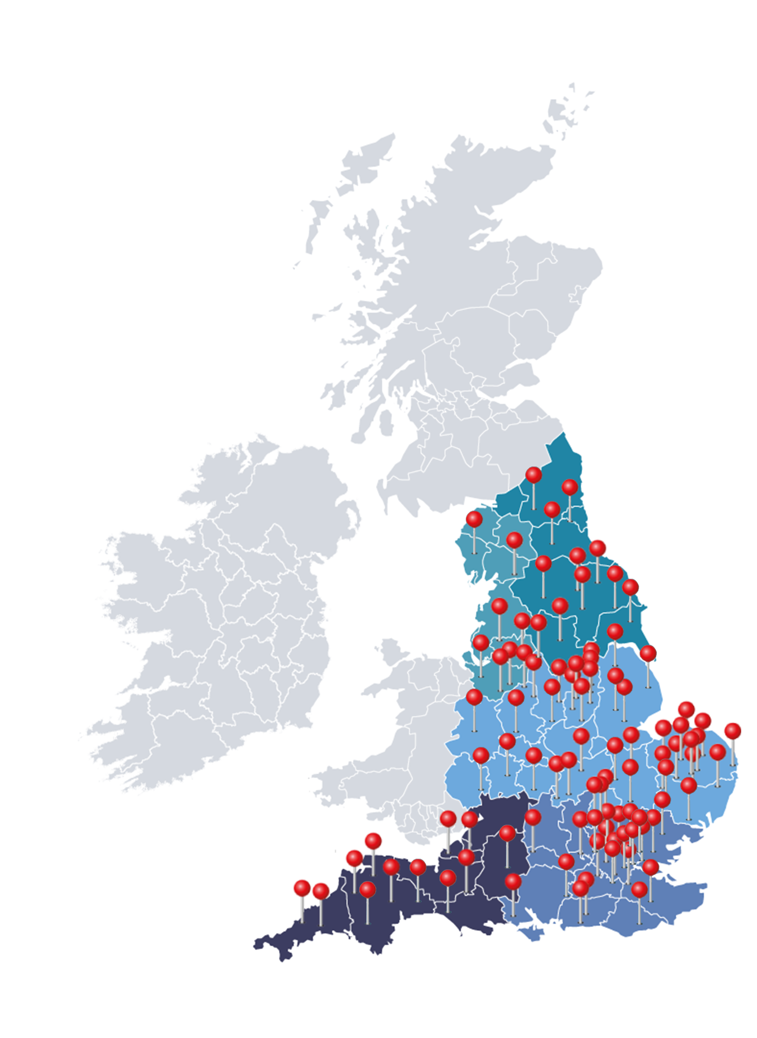
With PCN partnerships from Cornwall to Durham, Weston-Super-Mare to Southwold, our network of specialist physiotherapists operate face-to-face or remotely depending on your local needs.

Map of the UK