Go to Rehab Plans
Back to Conditions Pure FCP Service Expert FCPs for your Primary Care Network

Baker’s Cyst

What is a Baker's cyst?

  • A Baker’s cyst, also known as a popliteal cyst, is a swelling in the popliteal space (space behind the knee) that causes a visible lump.

How common is Baker's cyst?

  • Approximately 25% of patients with knee pain have a Baker’s cyst (1). However, this does not mean they are always the source of pain.
  • It is more common when a person has a knee condition such as osteoarthritis (2).
  • Approximately 5% of the population may have a Baker’s cyst in their lifetime (3).

Should I worry?

  • No.
  • Often, they are not the cause of knee pain and they can be incidental findings on scans (5).
  • A sudden increase in swelling, change of consistency, increased pain and/or altered sensation/weakness indicate a need for further specialist assessment (4).
  • If you have pain, swelling, changes to skin colour and temperature in the calf or thigh, this might be a sign of a serious condition called deep venous thrombosis which requires urgent medical attention.

Who is most likely to suffer from a Baker's cyst?

  • Those between 35 and 70 years of age (10).
  • Those with long-term knee conditions such as osteoarthritis.
  • Men and women are nearly equally affected (10).

What are the common symptoms?

  • Posterior (back) knee pain with an associated visible lump.
  • Limited range of movement with associated stiffness and tightness behind the knee.
  • A ruptured Baker’s cyst can cause sharp pain and redness in the calf (7).

What can I do?

  • Utilisation of anti-inflammatory medication which may be prescribed by your GP or pharmacist.
  • Regular icing over the affected region might help ease the pain.
  • Exercise and lifestyle adaptations to maintain strength and mobility of the knee.

How long will it take to recover?

  • Full recovery is dependent upon several variables including other underlying knee conditions.
  • Spontaneous resolution is common.
  • The visible lump might take a year or more to disappear. Often people will have a visible lump without pain.

1. Introduction

A Baker’s cyst (also known as a popliteal cyst) is swelling in the popliteal fossa (region at the back of the knee) which can lead to pain and stiffness in the knee (2). The terminology is somewhat misleading as technically it is not a true cyst. It originates from knee joint effusion (swelling) where fluid distends from the gastrocnemio-semimembranosus bursa (a fluid-filled sac between calf and hamstring muscles). It is more common in older people as part of a chronic knee condition, where there may be general swelling of the knee secondary to age-related degenerative changes (2).

There are both primary and secondary cysts. Primary cysts are usually asymptomatic (do not cause pain) and occur in younger patients. These cysts do not directly involve the knee joint. Secondary cysts are more common and directly involve the knee joint. They are more common in older people and are thought to be caused by muscular weakness around the knee and changes within the joint.

pure

2. Signs & Symptoms

Symptoms are typically localised to the back of the knee. These can include:

3. Causes

Any condition of the knee which causes degenerative changes or inflammation can contribute to the development of a Baker’s cyst. It is a common finding with intra-articular (within a joint) conditions including (3):

  • Meniscal tears (most common).
  • Anterior cruciate ligament deficiencies.
  • Cartilage degeneration.
pure

4. Risk Factors

This is not an exhaustive list. These factors could increase the likelihood of someone developing a Baker’s cyst. It does not mean everyone with these risk factors will develop symptoms.

5. Prevalence

Approximately 5% of the population may have a Baker’s cyst (3) and up to 25% of those with knee pain may have a Baker’s cyst on an ultrasound scan although this does not mean symptoms will exist (1). Men and women are nearly equally affected (10).

6. Assessment & Diagnosis

Musculoskeletal physiotherapists or other suitably qualified musculoskeletal specialists can provide you with an accurate and timely diagnosis by obtaining a detailed history of your symptoms. A series of physical tests might be performed as part of your assessment to rule out other potentially involved structures and gain a greater understanding of your physical abilities to help facilitate an accurate working diagnosis.

Imaging is not usually required to make a Baker’s cyst diagnosis, but your treating clinician may request further imaging if deep vein thrombosis or significant intra-articular pathology needs to be excluded. Patients with a Baker’s cyst and calf swelling should be referred urgently for appropriate imaging studies to exclude deep vein thrombosis (8,9).

7. Self-Management

As part of your treatment, your treating clinician will help you understand the condition and what needs to be implemented to effectively manage your symptoms. On-going treatment might not be necessary if you have an isolated, asymptomatic Baker’s cyst.

If you have an underlying condition that is causing the Baker’s cyst and it is painful, this may require further management. Over-the-counter pain relief and the application of ice may be used to reduce swelling and manage any pain. Activity modification strategies and regular adherence to a condition-specific rehabilitation programme should also form part of the management. It should be noted that rehabilitation exercises are not always a quick fix but, if adhered to on a consistent basis (weeks to months), over time they have been shown to yield positive outcomes.

pure

8. Rehabilitation

Your musculoskeletal physiotherapist may advise on a condition-specific exercise programme that is tailored to address any muscular weakness or imbalances, as well as ensuring the knee joint has an optimal range of movement. A more general approach to lifestyle changes and self-management of chronic conditions may also be addressed in your consultation.

9. Baker's Cyst Rehabilitation Plan

Treatment plan

This programme focuses on ensuring you achieve an optimal range of movement and good muscle strength. We suggest you perform this once a day for approximately 2-6 weeks as your symptoms allow. The exercises are unlikely to be painful but if there is any discomfort is should not exceed any more than 4/10 on your perceived pain scale.

Treatment plan  - Rating

10. Return to Sport/Normal Life

For patients wanting to achieve a high level of function or return to sport, we would encourage a consultation with a musculoskeletal physiotherapist as you will likely require further progression beyond the intermediate rehabilitation stage. Before returning to sport, a rehabilitation programme should incorporate plyometric-based exercises; this might include things like bounding, cutting, and sprinting exercises (5,7).

As part of a multi-modal treatment approach, your treating clinician may also use a variety of other pain-relieving treatments to support symptom relief and recovery. Whilst recovering you might benefit from a further assessment to ensure you are making progress and establish the appropriate progression of treatment.  Ongoing support and advice will allow you to self-manage and prevent future reoccurrence.

11. Other Treatment Options

  • Deteriorating and symptomatic Baker’s cysts which do not improve with conservative management may require surgical interventions. The main aim of surgery is to resolve any underlying conditions such as meniscal tears or ligament damage. Surgery aimed at treating underlying conditions can result in the resolution of the cyst. Interestingly, cyst excision (removal) is associated with a high rate of recurrence.
  • Deteriorating and symptomatic Baker’s cysts which do not improve with self-management may require aspiration (needle being inserted into the cyst and the fluid is drained out). This would require a referral to a medical specialist.

References

  1. Picerno, V., Filippou, G., Bertoldi, I., Adinolfi, A., Di Sabatino, V., Galeazzi, M. and Frediani, B. Prevalence of Baker’s cyst in patients with knee pain: an ultrasonographic study. Reumatismo (2013) , 264-270.
  2. Cao, Y., Jones, G., Han, W., Antony, B., Wang, X., Cicuttini, F. and Ding, C. (2014). Popliteal cysts and subgastrocnemius bursitis are associated with knee symptoms and structural abnormalities in older adults: a cross-sectional study. Arthritis research & therapy, 16(2), 1-9.
  3. Demange, M. K. (2015). Baker’s cyst. Rev Bras Ortop, 46(6), 630-633.
  4. Fritschy, D., Fasel, J., Imbert, J.C., Bianchi, S., Verdonk, R. and Wirth, C.J. (2006). The popliteal cyst. Knee Surgery, Sports Traumatology, Arthroscopy, 14(7), 623-628.
  5. Raghupathi, A.K. and Shetty, A. (2013). Unusual presentation of popliteal soft tissue sarcoma: not every swelling in the knee is a Baker’s cyst. Journal of surgical case reports, (10).
  6. Herman, A.M. and Marzo, J.M. (2014). Popliteal cysts: a current review. Orthopedics, 37(8), 678-684.
  7. Kornaat, P.R., Bloem, J.L., Ceulemans, R.Y., Riyazi, N., Rosendaal, F.R., Nelissen, Kloppenburg, M. (2006). Osteoarthritis of the knee: association between clinical features and MR imaging findings. Radiology, 239(3), 811-817.
  8. Kim, J.S., Lim, S.H., Hong, B.Y. and Park, S.Y. (2014). Ruptured popliteal cyst diagnosed by ultrasound before evaluation for deep vein thrombosis. Annals of rehabilitation medicine, 38(6), 843.
  9. Handy, J.R. (2001). Popliteal cysts in adults: a review. Seminars in Arthritis and Rheumatism 31(2), 108-18.
  10. Chatzopoulos, D., Moralidis, E., Markou, P., Makris, V. and Arsos, G. (2008). Baker’s cysts in knees with chronic osteoarthritic pain: a clinical, ultrasonographic, radiographic and scintigraphic evaluation. Rheumatology international, 29(2), 141-146.
Other Conditions in Knees, Lower Legs

Meniscus Injury

Structural knee injury, triggered either by a tear or through wear and tear.

Share on Social Media

Degenerative Meniscus

Seen to be normal as we age, but in some situations can result in knee aches, pain or joint swelling.

Share on Social Media

Patellar Tendinopathy

Knee pain at the lower border of the kneecap which is also known as ‘jumper’s knee’.

Share on Social Media

Fat Pad Impingement

A rare condition affecting the adipose (fat) tissue that sits under the kneecap (patella) between the joint spaces of the knee.

Share on Social Media

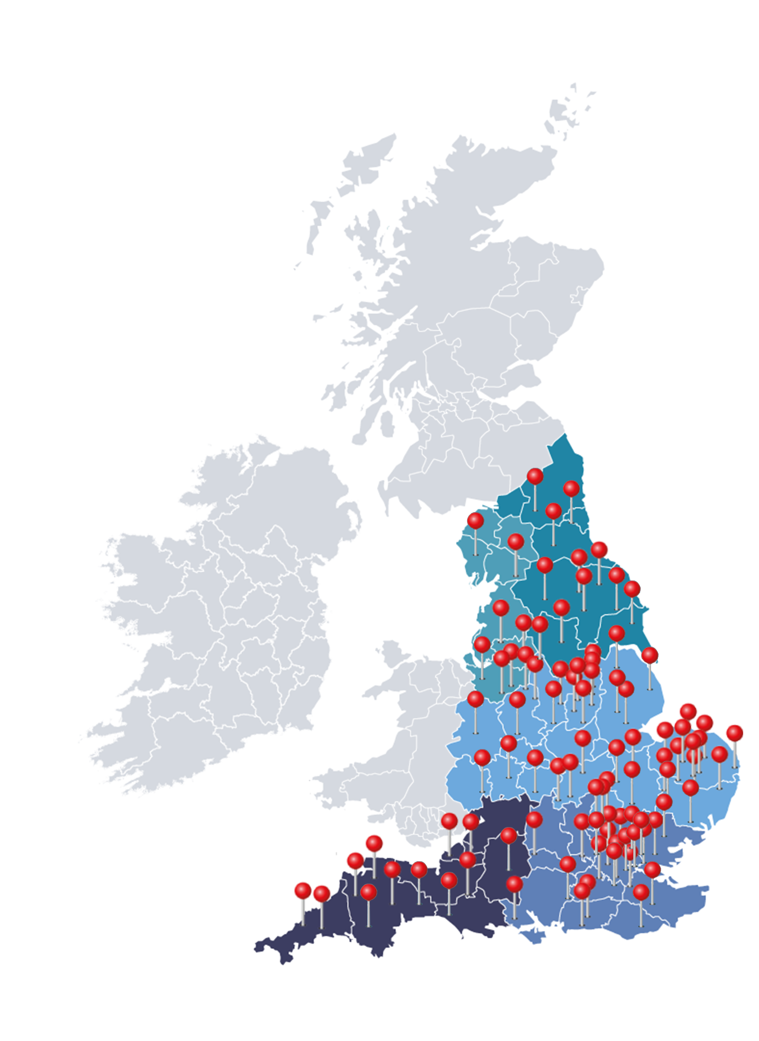
Our award-winning FCPs support PCNs and GP Federations all over England.

With PCN partnerships from Cornwall to Durham, Weston-Super-Mare to Southwold, our network of specialist physiotherapists operate face-to-face or remotely depending on your local needs.

Map of the UK